Macular Degeneration
Age-related macular degeneration, often referred to as AMD, is a medical condition that usually affects older adults. This vision-stealing disease is the result of degeneration to the macula. It results in a loss of vision in the center of the visual field because of the damage to the retina. It occurs in dry and wet forms and is the leading cause of blindness and visual impairment in adults over the age of 50.
Types of Macular Degeneration
Macular degeneration can make it difficult or impossible to read or recognize faces, although enough peripheral vision remains to allow other activities of daily life. The dry form of advanced AMD results from atrophy of the retinal pigment epithelial layer below the retina. This causes vision loss due to the damage of photoreceptors, also known as rods and cones, in the central part of the eye.
The wet form of advanced AMD causes vision loss due to abnormal blood vessel growth. This ultimately leads to blood and protein leakage below the macula. Bleeding, leaking, and scarring from these blood vessels eventually causes irreversible damage to the photoreceptors and rapid vision loss if left untreated. Fortunately, only about 10 percent of patients suffering from macular degeneration have the “wet” type.
Macular degeneration is not painful, which may allow it to go unnoticed for some time. For this reason, regular eye examinations are important. While approximately 10 percent of patients age 66 to 74 will have findings of macular degeneration, the prevalence increases to 30 percent for patients age 75 to 85 years of age. Family history may also play a factor. The good news is that regular eye exams, early detection, and new treatment options enable doctors to maintain (and in some cases increase) visual acuity in patients.
Watch our video to learn more!
The Amsler Grid (for Macular Degeneration)
The Amsler grid was developed by Marc Amsler, a Swiss ophthalmologist, and has been used since 1945. It is a grid of horizontal and vertical lines used to monitor a person’s central visual field. It is a diagnostic tool that aids in the detection of visual disturbances caused by changes in the retina, particularly the macula, such as age-related macular degeneration.
If you are at risk for macular degeneration, you can also use this chart at home to monitor your vision. Regular use of this chart, along with an annual comprehensive eye exam, provides you the best chance at accurate and early detection of macular damage. Early diagnosis means early treatment, and early treatment may help to limit or slow the progression of vision loss.
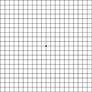
How to Test Yourself with the Amsler Grid
- If you need reading glasses, please wear them while you use the Amsler grid.
- The grid should be placed at reading distance from your eyes.
- Test your vision with adequate lighting.
- Test only one eye at a time. Cover the other eye.
- Focus on the dot at the center of the chart.
- Do any of the lines look wavy, blurred or distorted? (All lines should be straight, all intersections should form right angles and all the squares should be the same size.)
- Are there any missing areas or dark areas in the grid?
- Can you see all corners and sides of the grid?
It is VERY IMPORTANT to immediately contact your eye doctor to report any irregularities.
You can mark areas of the chart that are not seen properly and bring it to your eye exam.
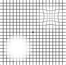
Examples of abnormal Amsler Grid tests:
Refer to the Patient Resources section of our website for a link to both an online and printable version of the Amsler grid.